


Since about 1990 with the introduction of TEACH at State Farm and Colossus at Allstate automobile insurance companies have worked off two basic formulas. The one that personal injury attorneys are all too familiar with is deny, delay, defendant. The other is the one most attorneys would do well to fully understand.
When James Mathis created TEACH at State Farm and managed Colossus for Allstate it was based on some very simple premises. His first question was what are we paying? This forced healthcare providers to become more proficient at diagnosing injuries, a skill that most doctors have not mastered.
This lack of diagnoses usually leads to a low-ball settlement offer. I use the plural because medical doctors tend to diagnose a cervical strain while chiropractors go with cervical sprain when dealing with whiplash type injuries. These two oversimplifications reduce the value of a claim. More importantly, they omit the more serious underlying injuries.

Mathis’ second question asked us to prove the diagnosis. By simply posing this question we now needed to have testing, reproducible objective testing to validate that diagnosis. What test do you have to verify your diagnosis, your clinical impression, your opinion?
For attorneys, opinions are what is all about. Two attorneys will present their opinions based on facts in and the court will render an opinion. Doctors may have plenty of opinions but clinically we need a diagnosis. From the Greek root of gnosis, to know, a diagnosis is something that a doctor knows about the patient’s condition.
This knowledge can be based in experience of having seen X number of cases with a similar set of symptoms and findings that an experienced doctor will know what to look for. Even with this experience, if the case gets to litigation you need proof of what you think you know.
For all the orthopedic/neurological testing that can be done the one test that is the most objective and most verifiable is radiology. Radiology offers an image of where injured structures are at a given time. Like the old adage of a picture being worth a thousand words, in a personal-injury case these pictures can be worth thousands of dollars.
As with any testing there is variability and especially variability of interpretation but for the trained eye especially one with a digital ruler with which to measure, these images become irrefutable.
As with any testing there is variability and especially variability of interpretation but for the trained eye especially one with a digital ruler with which to measure, these images become irrefutable. Usually, when an attorney asks me to review a file I will receive copies of the radiology reports. Sometimes that alone is all the information I need. When the written information is insufficient, viewing and measuring the films myself may reveal more serious injuries that may be the cause of permanent impairment.
So what can be found on radiology. With plain film X-rays misalignment may lead to additional testing. Fractures, often very small and compression fractures can add considerable value to a case.
When the plain film study does not provide enough information to explain the patient’s complaints an MRI may be in order. These studies of the spine not only show a more detailed picture of the bony structures but also involvement of injured discs that are more serious cause of pain.
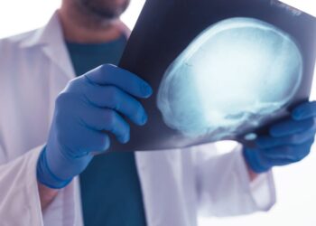
When on the subject of MRIs, it is also important to consider brain scans. In attempting to detect lesions in the cerebral cortex a stronger magnet, 3T will usually reveal far more findings than the weaker 1T MRI. A multitude of other studies performed with this MRI can reveal brain injuries that might otherwise go unproven.
In addition, a stand-up MRI offers two advantages. First, the additional weight on the discs in this position will cause weakened discs to bulge farther then they would on a recumbent MRI. This simple procedure will demonstrate far more disc injuries than a recumbent MRI. The weaker magnet on an upright or weight bearing MRI makes it less desirable when studying brain injuries with one exception. This goes to the cerebellar tonsils which after a hyperextension/ hyperflexion trauma can cause them to fall into the foramen magnum causing myriad symptoms.
With a recumbent MRI the brain will slip back into the cranium and this problem can often go undetected. With the patient upright the structures will fall toward the foramen magnum and detection of this injury increases three-fold. Bill Gallagher